Author Archives: petscans101


Purpose and Application of PET Scans

PET scans, Positron Emission Tomography scans, are a type of nuclear medicine imaging. It is a medical imaging system which helps doctors detect illnesses in patients. Its purpose is to show the functioning and structures of tissues and organs. In the medical field, these scans are used to provide information on the shape and size of the parts of the body and can determine flow of blood, the usage of oxygen and sugar/glucose metabolism. A PET scan is very useful when related to sugar metabolism because it can show it before it is physically visible. PET scans are mostly used to detect cancer in tissues or to examine the effects of cancer treatments. It is also used to detect heart disease such as coronary artery disease and brain illnesses such as Alzheimer’s disease and epilepsy. All information shown by the PET scan can be used to diagnose illnesses and to make decisions about treatment. The advantage of PET scanning is that it may show more wide-ranging diseases than CT (computed tomography) or MRI (magnetic reasoning imaging) scans. This type of scan can also be combined with a CT scan to produce one, single image. When it is fused with a CT scan, it gives more precise data about the body.


(BY: Chinthuri Selvarajah)
How Does a PET Scan Work?

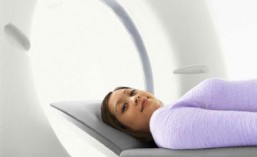
The PET scanning system is not dangerous and is harmless. PET scans effectively work by using a radioactive glucose injection. Firstly, a radioactive material called a radio tracer that’s combined with sugar, FDG (18-Fluorodeoxyglucose), is injected into the patient’s bloodstream or in some cases, swallowed or inhaled. The organs that are affected will process the FDG and as a result, the PET Scans will detect the radiation from those organs in the patient. This is because, the radio tracer in the body will be releasing positrons (positively charged particles) that the scanning system will read. The computer will then collect the information on the area where the positrons are and it translates it into two- or three- dimensional images of the body’s tissues and organs. These images can be combined with CT scan images to form ultimately one overall image. If anything is detected, further precautions will need to be taken and the PET Scans can be used for diagnosis, evaluation and treatment of disease.

(BY: Samantha Charles)
Revolving PET Scan

Scan Analysis #2

These series of images show the development of Alzheimer’s disease. The left image is a PET scan that shows the normal levels of glucose metabolism (red and yellow) in the brain. As you can see, the levels of glucose metabolism are decreased in the second and third PET scan. The middle image shows the glucose metabolism decreased however the brain is still full. It is only a mild infection at the moment. This is when the patient may begin to forget things, that may seem normal to forget, however could progress into more severe memory loss. Finally, the last image shows the patient’s brain as it has much less glucose metabolism than it did when it was normal. Also, if you noticed, now the scan has a big, black empty hole in the middle of the brain image than when it started. This is because the disease has gotten to the point where the patient isn’t able to remember very much. The development of the brain at this point is very low.
(BY: Samantha Charles)
Scan analysis

This PET scan is showing the difference in brain images. As you can see from the picture there are 4 different brain images all with difference pH readings. The top, left image has a pH of 18. The top, right image has a pH of 19. The bottom, left has a pH of 24, and the bottom, right image has a pH of 25. These PET scans are used to show the amount of sugar compared to the acidity of the scans. The top, left image has the lowest pH and also has the least amount of sugar (represented by the yellow). The bottom, right image has the most yellow, as well as some green, which shows the severity of the amount of sugar. So you can see that as the pH is increased, the sugar is increased as well. As a result, this patient has medically uncontrollable seizures which are also known as epilepsy.
(BY: Samantha Charles)
Ethical Issues Regarding PET Scans

There are a couple of ethical issues regarding the use of PET scans. Firstly, PET scans are occasionally inaccurate and as a result may give false images of the body. One ethical issue that comes up because of this is that people wonder if it is right to use this technology when it could possibly give misconceptions and can have a great impact on the patient. This scanning system can misdiagnose a person which would not only cost the doctors who are treating the “sick” patient but the patient themselves. It would waste time and effort and can be avoided by not using the scanning system for mild issues. For example, if a woman got a PET scan done and it showed that she “had” cancer when she really didn’t she would have to go through days of treatment. This would not only affect the person physically but also mentally. The family and loved ones of that person will also be part of this issue because cancer is a very sensitive topic. Also, patients that actually have a disease may not get the needed scans and treatment done due to the fact that the doctors are busy treating patients that have been misdiagnosed. Clearly, the fact that PET scans can be inaccurate may cause many problems.
Secondly, to get a PET scan done, you must be injected by radioisotopes. This is a radioactive material that contains radiation. As known by many, radiation is one of the many causes of cancer. Ironically, PET scans can possibly cause cancer (not proven as of yet) and can diagnose cancer. There is an ethical problem because it is not known if it is right to be using these types of materials which have the ability to damage the health of our bodies. Some argue that the radiation is in small amounts and it does not stay in the body for long. However, these types of medical facts can be proven wrong and therefore we can never be sure about the impacts of this scan. Also, everybody is different, so some bodies may hold the radioisotopes in the body longer and others may not. This can clearly affect the body negatively since the chemical was not meant to stay in the body for a long time. Another problem is that weaker bodies may not be able to handle the radiation as much as stronger bodies. This can also affect how the body reacts to the radioisotopes and may cause problems within the body of the patient. Obviously, the radioisotopes bring up many ethical questions about the safety of the use of PET scans.
In conclusion, although there are several ethical issues, PET scans can also allow doctors to observe detailed views of the body.
(BY: Chinthuri Selvarajah)
Bibliography
Images:
Alzheimer’s scan. (2009, August 26). [Photograph]. Retrieved November 2, 2012 from http://www.triumf.ca/research-highlights/experimental-result/cutting-edge-alzheimers-research
Brain Wellness Innovators. (n.d.). [Photograph]. Retrieved October 31, 2012 from http://www.jungsoul.com/HealYourBrain/home.html
How A PET Scan Works. (2008). [Photograph]. Retrieved October 29, 2012 from http://micpetct.com/howpetworks.htm
How the PET scan works. (2004, August). [Photograph]. Retrieved November 4, 2012 from http://www.scq.ubc.ca/looking-inside-the-human-body-using-positrons/
Positron Emission Tomography Scanning-PET Scans. (n.d.). [Photograph]. Retrieved October 31, 2012 from http://healthinformatics.wikispaces.com/Positron+Emission+Tomography+Scanning+-+PET+Scan
(Sc)anticipation. (2011, June 26). [Photograph].Retrieved November 1, 2012 from http://sintelligently.blogspot.ca/2011_06_01_archive.html
Seizures. (2012). [Photograph]. Retrieved November 1, 2012 from http://www.mayfieldclinic.com/PE-PET.htm
The Equipment. (n.d.). [Photograph]. Retrieved November 1, 2012 from http://www.cyberphysics.co.uk/topics/radioact/PET.htm
The Unambiguous News. (2011, June 29). [Photograph]. Retrieved November 1, 2012 from http://sintelligently.blogspot.ca/2011_06_01_archive.html
What PET Sees. (2008). [Photograph]. Retrieved November 1, 2012 from http://micpetct.com/whatpetsees.htm
Why PET Works. (2008). [Photograph]. Retrieved October 31, 2012 from http://micpetct.com/whypet.htm
Books:
Jones, S. (2006). Imaging for Nurses. Oxford, UK: Wiley-Blackwell
Sandner, L., Ellis, C., Lacy, D., Little, C. & Mace, H.A. (2009). Investigating Science 10. Canada: Pearson Canada, Inc.
Websites:
Diagnostic Imaging Center at NYU Clinical Cancer Center. (November 15, 2010). What is PET/CT. Retrieved October 26, 2012 from http://www.med.nyu.edu/di/petCt/faqs.html
How Stuff Works. (2000, October 18). How Nuclear Medicine Works. Retrieved October 29, 2012 from http://www.howstuffworks.com/nuclear-medicine1.htm
Mayfield Clinic. (2010). PET (positron emission tomography) scan. Retrieved November 1, 2012 from http://www.mayfieldclinic.com/PE-PET.htm
PET Scans Ontario. (n.d.). Explaining PET Scans. Retrieved October 26, 2012 from https://www.petscansontario.ca/cms/One.aspx?portalId=69866&pageId=69880
Society of Nuclear Medicince and Molecular Imaging. (2012). Positron Emission Tomography (PET) Scan. Retrieved November 1, 2012 from http://www.molecularimagingcenter.org/index.cfm?PageID=7608
